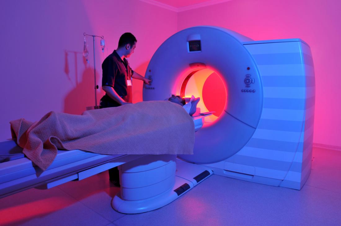
MRI (Magnetic Resonance Imaging) scanners are crucial diagnostic tools that allow doctors to examine the internal structures of the human body without the need for invasive procedures. The process of manufacturing these complex devices involves highly sophisticated technology and expert craftsmanship to ensure precise imaging capabilities. From superconducting magnets to powerful computer systems, each component is designed and assembled with meticulous care. Understanding how MRI scanners are made can give insight into the innovation behind the advanced healthcare solutions that save lives daily. This blog will walk you through the key stages involved in the manufacturing of MRI scanners, highlighting the major components and processes behind this life-saving technology.

The Role of Superconducting Magnets
At the heart of every MRI scanner lies the superconducting magnet, which generates the powerful magnetic field essential for the imaging process. These magnets are created using coils of superconducting wire that must be cooled to extremely low temperatures using liquid helium. This process ensures that the wire can conduct electricity with zero resistance, allowing the magnet to generate a stable magnetic field. The strength of the magnet plays a significant role in the resolution of the images, with stronger magnets providing more detailed scans. Superconducting magnets typically have magnetic field strengths of 1.5 Tesla or higher, with some advanced MRI machines using magnets that exceed 3 Tesla.
Manufacturing the Gradient Coils
The gradient coils are another critical component of MRI scanners, responsible for producing the varying magnetic fields necessary to create detailed images of different body parts. These coils are typically made from copper wire and are designed to work in conjunction with the main superconducting magnet. Gradient coils help to localize the magnetic field, allowing the scanner to focus on specific regions of the body. Their design is particularly complex, as they must be able to generate rapidly changing magnetic fields without distorting the overall image quality. Precision in the construction of gradient coils is crucial for ensuring high-quality imaging performance.
The RF Coils and Signal Reception
Another essential component of MRI scanners is the radiofrequency (RF) coils, which are responsible for transmitting and receiving signals during the scanning process. These coils emit RF pulses that interact with the body’s hydrogen atoms, which then emit signals that are detected by the same coils. RF coils are tuned to specific frequencies to target particular tissues, ensuring that the MRI scanner can obtain the most accurate data possible. The signals picked up by the RF coils are then processed by the system’s computer to generate detailed images. The design of these coils is vital for ensuring clarity and resolution in the final scan.
The Role of the Computer System
The computer system within the MRI scanner acts as the brain of the machine, processing and interpreting the data gathered by the RF coils. It takes the raw signals and transforms them into images that doctors can use to diagnose a wide variety of medical conditions. The system utilizes complex algorithms and image reconstruction techniques to enhance image clarity and accuracy. Modern MRI systems employ advanced software to correct distortions and improve the final output. This computational power is one of the key reasons why MRI scans are so precise and valuable in medical diagnostics.
Cryogenic Cooling Systems
To maintain the low temperatures necessary for superconducting magnets, MRI scanners are equipped with cryogenic cooling systems, typically using liquid helium. These cooling systems are vital for ensuring that the superconducting magnets remain at the required temperature for optimal performance. These systems must be carefully monitored and maintained to prevent any temperature fluctuations that could impact the scanner’s functionality. The cooling process requires a significant amount of energy and infrastructure, making it one of the more expensive aspects of MRI manufacturing. The use of liquid helium also raises concerns about sustainability and supply, leading to ongoing research into alternative cooling methods.
Vote
Who is your all-time favorite president?
MRI Scanner Housing and Safety
The outer housing of an MRI scanner is designed to provide both structural integrity and patient safety. The scanner is typically encased in a robust frame made of materials like steel or aluminum to protect the sensitive components inside. The housing also ensures that the powerful magnetic field does not interfere with surrounding electronics, which could disrupt the scanning process. Additionally, the design of the housing allows for easy access to the patient while also ensuring their comfort during the scan. Strict safety guidelines are followed in the design and construction of MRI scanner housing to protect both the patients and the medical staff.
The Manufacturing of the Patient Table
The patient table is another key part of the MRI scanner that requires precise engineering. This table must be capable of moving smoothly and accurately into the MRI machine, positioning the patient in the right spot for optimal imaging. Typically, these tables are made of non-ferromagnetic materials, such as carbon fiber, to prevent interference with the magnetic field. The design also prioritizes comfort, as patients may need to lie still for extended periods during the scan. Customizable tables are available for specific procedures, allowing the machine to adapt to different types of diagnostic imaging needs.
Assembly of MRI Components
The assembly of all the components into a fully functional MRI scanner is a meticulous process that involves careful alignment and calibration. Each part—whether it’s the superconducting magnets, gradient coils, RF coils, or computer system—must be installed and calibrated to ensure the machine works seamlessly. The assembly process also includes testing each component to ensure it meets rigorous quality control standards. After assembly, the machine undergoes comprehensive testing to ensure that it delivers clear, high-resolution images without errors. This process is crucial to making sure the MRI scanner meets medical standards.
Advertisement
Testing and Calibration
Once the MRI scanner is fully assembled, it must undergo a series of tests to ensure that it operates at its full potential. This includes testing the magnetic field strength, verifying image quality, and calibrating the RF coils to ensure proper signal reception. Testing is a critical step that ensures the scanner can deliver consistent and reliable results. During this phase, technicians will also check the cooling system, gradient coils, and other components to ensure that everything is functioning correctly. Only after passing these tests is the MRI scanner ready for clinical use.
The Future of MRI Scanner Manufacturing
With advances in technology, the future of MRI scanner manufacturing looks promising. New developments in materials science and computational power are enabling the creation of more compact and efficient MRI machines that offer even better image quality and faster scan times. Researchers are exploring the use of high-temperature superconductors that could eliminate the need for liquid helium, reducing costs and environmental impact. As MRI technology continues to evolve, we can expect to see machines that are not only more powerful but also more accessible and affordable. The future of MRI scanner manufacturing is set to revolutionize the way healthcare providers diagnose and treat a wide variety of conditions.
Key Steps in MRI Scanner Manufacturing
- Superconducting magnets are designed and manufactured using coils of superconducting wire.
- Gradient coils are created to produce varying magnetic fields for precise imaging.
- RF coils are carefully designed to transmit and receive signals from the body’s hydrogen atoms.
- The computer system processes and interprets the data to generate high-resolution images.
- Cryogenic cooling systems are integrated to maintain the temperature of the superconducting magnets.
- MRI scanner housing is built for structural integrity, safety, and patient comfort.
- The patient table is manufactured with non-ferromagnetic materials for smooth movement.
Important Components of MRI Scanners
- Superconducting magnets
- Gradient coils
- RF coils
- Computer system for image processing
- Cryogenic cooling systems
- Scanner housing
- Patient table
Pro Tip: When using an MRI scanner, always follow the technician’s instructions to ensure you are positioned correctly for the most accurate scan results.
| Component | Purpose | Material |
|---|---|---|
| Superconducting magnets | Generates the magnetic field | Coils of superconducting wire |
| Gradient coils | Generates varying magnetic fields | Copper wire |
| RF coils | Transmits and receives signals | Custom-designed materials |
“The evolution of MRI technology continues to transform the healthcare industry, enabling more accurate diagnoses and better patient outcomes.”
The manufacturing process behind MRI scanners involves a delicate balance of precision engineering, innovation, and safety protocols. From superconducting magnets to advanced cooling systems, every component plays a critical role in ensuring the success of the scanner. As technology advances, the manufacturing of MRI scanners is becoming more efficient and environmentally friendly. Whether you are a healthcare provider or a patient, understanding how MRI scanners are made can help you appreciate the cutting-edge technology behind the machine. Share this information with your network and bookmark it for future reference, as understanding MRI scanner manufacturing can lead to a deeper appreciation of medical imaging advancements.